Seattle Neurology
  • Home
  • Blog
  • EBook
    • Migraine
    • Polyneuropathy
    • Low Back Pain
  • Resources
    • Conditions >
      • Aphasia
      • Brain Trauma
      • Brain Tumor
      • Dementia >
        • Alzheimer's Disease
        • Frontotemporal Dementia
        • Vascular Dementia
      • Diabetes
      • Dystonia
      • Epilepsy
      • Headaches
      • Hearing Loss
      • Low Back Pain
      • Multiple Sclerosis
      • Parkinson's Disease
      • Polyneuropathy
      • Postural Tremor
      • Sleep Disorder
      • Stroke
      • Vision Impairment
    • Geriatric Issues >
      • Agencies
      • Caregiving
      • End of Life
      • Independent Living Facility
      • Medical Supplies
      • Social Services
    • Other >
      • Disability
      • General Health
      • Pharmaceuticals
      • Rehabilitation

CASE STUDY #11

5/2/2014

0 Comments

 
An 83 y/o man was out for a daily walk in his neighborhood when he was discovered by a passerby to be face-down bleeding from his nose.  He was rushed to the local ED where he was confused and complaining of pain at multiple sites.  After extensive tests, the only pertinent abnormality was a small bleed in the lateral ventricle of the right frontal lobe of his brain.  Once returned to home and healed from his minor bruises and strains, it was clear to his family that there had been a change to this patient both physically and emotionally.  He no longer walked confidently and now exhibited small cautious steps.  For the first time in his life, he began having problems with urinary urgency in which he would have to hurry to the bathroom in order to urinate.   At night he began wearing adult diapers.  He also became more confused and had to be looked after by his wife.  In essence, he regressed to being more like a toddler.  His wife had to assume more of the daily household activities.   He stopped driving.  
On exam, the most striking feature was a slow, cautious gait with difficulty turning around.  Otherwise the exam was non-focal. 

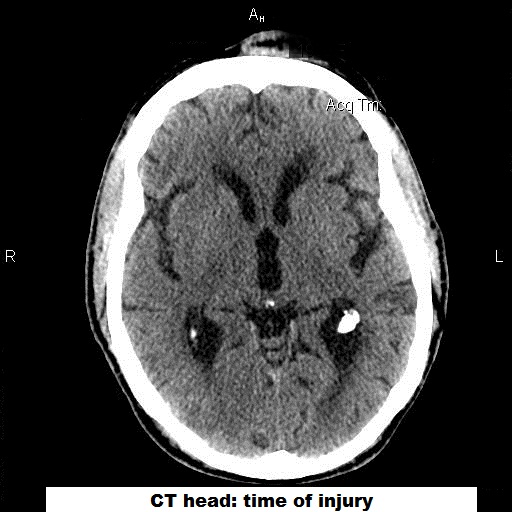
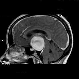
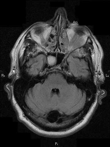
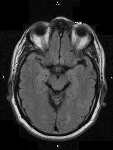
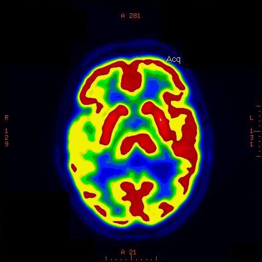
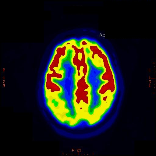
First, an MRI of the brain showed signs that cerebrospinal fluid (CSF) was pushing into the adjacent brain (‘transependymal flow’).  In addition, when comparing the size of the ventricles (the chambers found in the center of the brain that house the CSF) to the size of the ventricles from a pre-accident MRI, it was clear that the ventricles were much larger.   This suggested that he had acquired hydrocephalus (‘water on the brain’). 

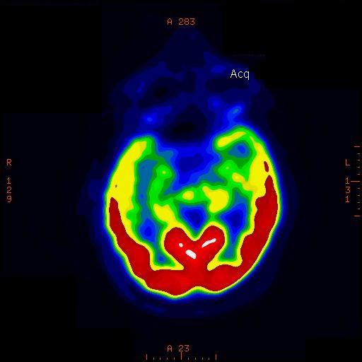
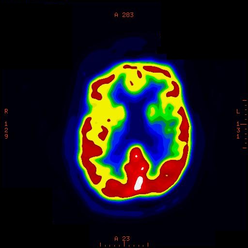
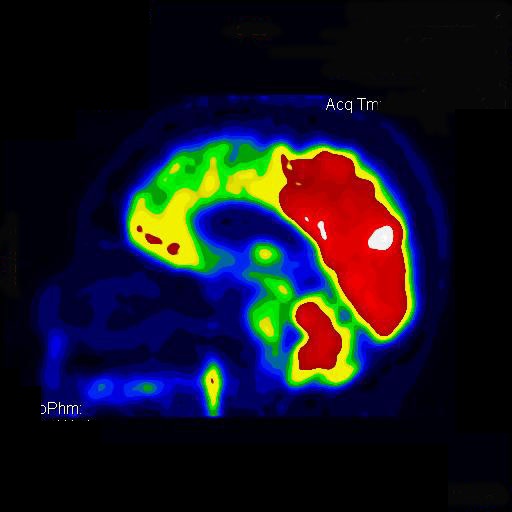
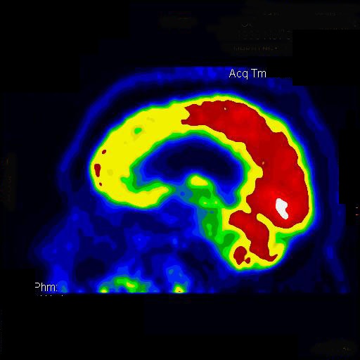
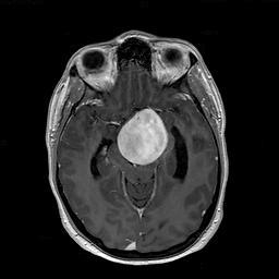
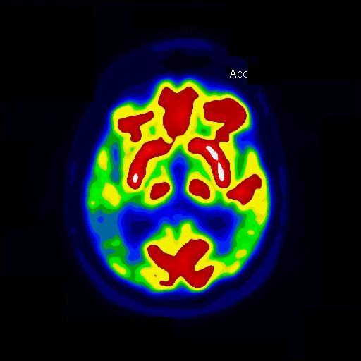
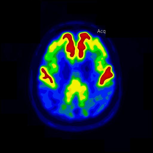
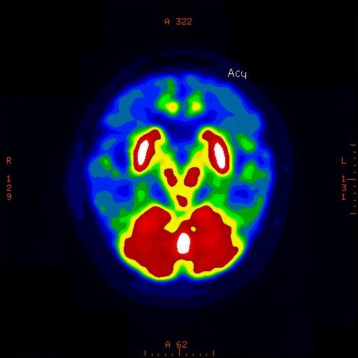
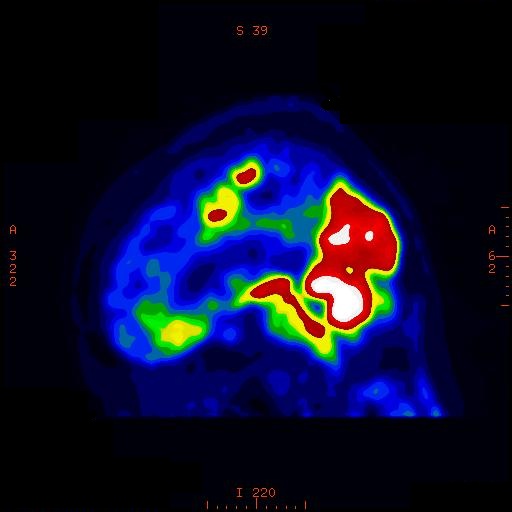
I then sent him for a lumbar puncture which revealed a normal pressure when performed – rather than an elevated pressure as one might expect.  I then performed a nuclear study called an Indium-111 cisternogram in which radioactive Indium-111 is injected into the CSF around the lumbar spinal cord.  In a normal person, this Indium-111 will not collect inside the ventricles but with patients who have a condition called Normal Pressure Hydrocephalus (NPH), the Indium-111 does end up inside the lateral ventricles.  This test was positive for this patient.

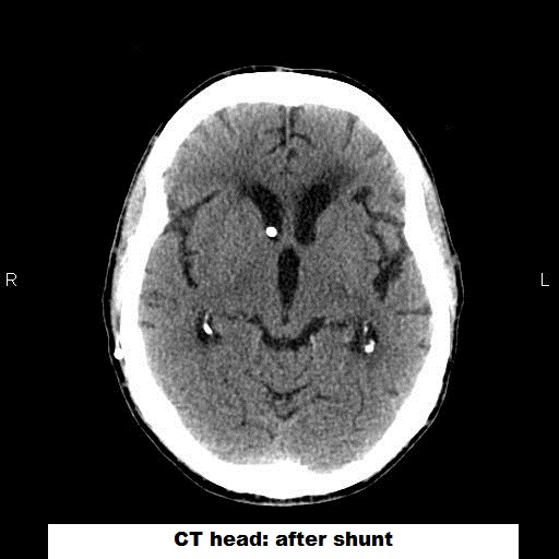
 He underwent placement of a ventriculoperitoneal (VP) shunt and immediately showed a return to his normal walking pattern and had resolution of his urinary urgency.  The confusion improved as well.  
DIAGNOSIS: NPH

Normal Pressure Hydrocephalus is thought to occur as a result of blockage of arachnoid granulations by degraded blood products.   Normally, the arachnoid granulations are responsible for resorption of the used CSF.  If an individual has experienced an intracranial bleed then they are theoretically at risk for developing NPH afterwards.  The classic clinical triad is ‘wild, wet and wobbly’ meaning worsening cognition, change in urinary habits and change in gait.  The treatment is placement of a VP shunt which will drain a small amount of the CSF continuously into the person’s gut.  
0 Comments

CASE STUDY #10

3/10/2014

0 Comments

 
This is a 32 y/o man who two weeks prior experienced the painless onset of tingling of both feet extending up to his calves.  He had no weakness.   One week later, he noticed that each time he hyperextended his neck there was a tingling pain that would shoot around from his back to his nipples and this would go away if he returned his neck to a normal position.  Four days later, he began having difficulty initiating a stream of urine and by this time his balance had deteriorated to the point that he was forced to clutch onto furniture just to walk in his condo.  On examination, he had weakness for hand grip of the right hand, inability to feel a vibrating tuning fork on the toes, loss of pin sensation all the way up to a point just between his shoulder blades (in the center of his back) and a wide base to his stance as he was very unsteady.    
The MRI of the cervical spine demonstrates congenital narrowing of the spinal canal.  In addition, there are relatively small disc bulges and protrusions at several levels which in combination with the congenital spinal stenosis results in injury of the cervical spinal cord.  
Picture
Picture
DIAGNOSIS: Acute Cervical Cord Compression from Cervical Spondylosis 
0 Comments

CASE STUDY #9

2/10/2014

0 Comments

 
This is a 68 y/o woman who for the prior four years showed a gradual worsening in her thinking that was evident to family members.  She had a change in her personality in which she became more disinhibited and careless.  For example, she began gambling at the casinos on a more regular basis losing up to $300 a month.  When her husband would confront her about her continual losses, she would respond, “Oh well – at least I’m having fun!”  Recently, her speech had grown slurred and she was having unexplained falls.  On exam, she demonstrated a wide-eyed expression on her face and could not move her eyes completely upward or downward.  She had a very rigid neck and there were brisk reflexes throughout all four limbs.  She had a tremor of her hands when she outstretched them in front of her body.  She could not draw the face of a clock correctly.

PET CT 
DIAGNOSIS:  Progressive Supranuclear Palsy (PSP)

Progressive Supranuclear Palsy (PSP) is diagnosed in 30 of 100,000 individuals each year.  For every 100 patients with Parkinson’s Disease, there is 1 person with PSP. 

PSP typically first presents during the 6th decade of life with problems walking.  Individuals will complain of unexplainable falls or stiffness often falling backwards instead of forwards.  The individual with PSP often develops insomnia as a part of the disease.  A small percentage will develop a rest tremor and be misdiagnosed as having Parkinson’s disease.  Later in the disease, they will have difficulty moving their eyes upward or downward completely causing them to spill their food when they eat or having difficulty descending stairs.  They will tend to decrease the amount of blinking which will lead to dry eyes.  The speech will become slurred making it difficult to understand what they are saying.  Much later there will be problems with their thinking resulting in a change in their personality as they become more forgetful, possibly cantankerous and indifferent.

This disease is a result of neuronal loss throughout the midbrain of the brainstem.  Later in the disease there will be significant neuronal loss within the parietal lobes and the regions of the frontal lobes responsible for initiating movements of the limbs.  The neurons demonstrate an accumulation of tau within the cell body prior to dying.  There is no known treatment to stop the progression of the disease.  There are medications to help ameliorate some of the symptoms.  The median interval from onset of the initial symptom to confinement in a wheelchair is 8 years.   
0 Comments

CASE STUDY #8

12/10/2013

0 Comments

 
This is a 34 y/o woman who over the past 6 months developed a clumsy right hand, loss of vision in her right peripheral field and slowing of her thinking.  During the past few weeks there had been some episodes of bladder urgency with some incontinence.  On exam, the right pupil was very slow to react to bright light from a flashlight.  She did not see as well in her right visual field.  The right side of her face was weak.  The right arm did not feel a pin as well as the left arm.   

This study demonstrated a large saccular aneurysm off the proximal intracranial left ICA.  This aneurysm is compressing the top of the left brainstem.  This part of the brainstem carries the impulses to and from the right side of the body (face, arm and leg). 

MRI brain with contrast
DIAGNOSIS: Left ICA Aneurysm 
0 Comments

CASE STUDY #6

9/20/2013

0 Comments

 
This is a 68 y/o man with a 4 month history of tingling of the left cheek with superimposed stabbing pain.  On exam, there is diminished pin sensation over the left cheek.  An initial MRI of his head was read as normal.

First, I performed a blink reflex in my office.  This is an electrical test in which I stimulate the trigeminal nerve and force an individual to blink.  I record how long it takes for a person to blink.  In this situation, there was a clear prolongation for blinking for the left lower eyelid relative to the right lower eyelid.  I then sent him for a detailed high-resolution MRI of the left eye.

This MRI scan shows a subtle lesion just under the left eyeball.  A biopsy of this tumor showed lymphoma.

MRI Brain
He was referred to an oncologist who treated him with systemic chemotherapy and he has been in remission for the past 6 years.  All of his facial symptoms resolved completely with treatment of the underlying lymphoma. 

DIAGNOSIS: 
Lymphoma 
0 Comments

CASE STUDY #5

7/15/2013

0 Comments

 
This is a 50 y/o woman who suffered a traumatic injury of her left peroneal nerve and shortly thereafter developed heightened sensitivity to light touch over the top of her left foot accompanied by a patch of burning pain as if she had experienced a sunburn on top of her foot.  The skin on top of her foot turned red and felt warmer relative to the rest of her foot.  
Picture
DIAGNOSIS: RSD 

Reflex Sympathetic Dystrophy (RSD) can develop following trauma to a region of the body.  Typical situations would be a severe ankle sprain, shoulder dislocation or fractured bone.  It can also occur with a stretch injury of a peripheral nerve.  RSD is also known as Complex Regional Pain Syndrome I (CRPS I).  Symptoms of RSD include …

a) motor impairment: weakness and dystonia
b) sensory abnormalities: burning pain (dysesthesia), hypersensitivity to touch (allodynia)
c) autonomic deregulation
    i) vasomotor: swelling (edema), discoloration (erythema, pale), temperature (warm, cold)
    ii) sudomotor: sweaty or dry (hyper/hypohydrosis) 
d) trophic changes: thinning of skin, loss of hair, nails turn brittle

This is one of the reasons that clinicians try to control the pain early following a trauma so that the patient can maintain a normal level of physical activity and hopefully avoid unnecessary long-term complications such as RSD.
0 Comments

CASE STUDY #4

3/13/2013

0 Comments

 
This is a 59 year-old man with a 4 year history of worsening short-term memory and difficulty expressing himself.  On initial exam, he misspoke frequently and made a number of spelling errors when asked to write a sentence.  He could not draw a clock. He was unable to recall 3 words after a short delay. He scored an 18/30 on the Mini-mental exam.  An MRI of the brain was normal.


This PET CT scan shows diminished blood flow in the parietal and temporal lobes bilaterally along with decreased flow to the posterior cingulate gyrus.  This is typical for Alzheimer’s disease. 

PET CT
DIAGNOSIS: Alzheimer’s Disease
0 Comments

CASE STUDY #3

1/14/2013

0 Comments

 
This is a 77 y/o retired woman with worsening problems expressing herself.  She also cannot troubleshoot how to fix her answering machine, drives slower than traffic and is forgetful.  On exam, she cannot recall 3 words after a short delay.  Her speech is halting and sub-fluent making it difficult for her to articulate her thoughts.   She cannot describe major news events.  She substitutes pronouns and vague terms such as 'that tool used to open the cans with' instead of 'can opener'.  The score on the mini-mental is 25/30 when two years earlier it has been 29/30.  An MRI of the brain performed two years earlier revealed signs of small-vessel disease which had been the result of years of untreated high blood pressure but no other abnormalities. 

This PET CT study demonstrates a focal region of decreased blood flow to only the posterior portion of the left hemisphere.  This fits with the vascular distribution for one of the major cerebral arteries.  This woman suffered an embolic stroke at some point since her prior MRI of the brain. 

PET CT
DIAGNOSIS: Left MCA Stroke 
0 Comments

CASE STUDY #2

12/12/2012

0 Comments

 
This is a 51 year-old woman who started having difficulty with her short-term memory at age 41.  She would frequently forget the names of people she met.  During the past five years, her husband has noticed that she repeats herself frequently – often asking the same questions.  On initial exam, there were no glaring abnormalities other than she could not repeat a list of 3 words that had been given to her to remember.  An MRI of the brain was normal. 

This PET CT study shows decreased blood flow to both hemispheres in the regions of the parietal and temporal lobes (right more than left).  This pattern is seen in early Alzheimer’s disease.


PET CT
DIAGNOSIS: Alzheimer’s Disease
0 Comments

CASE STUDY #1

5/20/2012

0 Comments

 
This is a 52 year-old woman who started having difficulty calculating her monthly bills beginning at age 42.  She developed personality changes, problems with short-term memory loss and even had difficulty reading a watch by age 46.  She had to stop working as a hairdresser and go on disability at age 48 and was placed in a nursing home by age 50.   On exam, she produced no intelligible speech and followed nothing but the most simple commands (‘stand’ or ‘sit’).  She paced about the room frequently seldom making eye contact.

This PET CT study demonstrated almost complete absence of blood flow to most of the surface of the brain with the exception of the very posterior portion of both hemispheres (occipital cortex).  This part of the brain is responsible for interpreting vision.  In addition, there are some ‘islands’ neurons deep in the white matter responsible for processing incoming sensory information called the thalamus.  This study shows that the thalami were healthy.  This is compatible with end-stage dementia.  It’s impossible to speculate at the underlying disease (Alzheimer’s, Pick’s Disease, Primary Progressive Aphasia, etc) based on this imaging study as the disease is too advanced. 


PET CT
DIAGNOSIS: End-stage Dementia
0 Comments

    Categories

    All
    Body Systems
    Book Review
    Case Study
    Dementia
    Diagnosis
    Elderly
    General Health
    Healthcare
    Helpful Tips
    Mental Health
    Migraines
    Parkinson's

    Archives

    May 2014
    April 2014
    March 2014
    February 2014
    January 2014
    December 2013
    November 2013
    October 2013
    September 2013
    August 2013
    July 2013
    June 2013
    May 2013
    April 2013
    March 2013
    February 2013
    January 2013
    December 2012
    November 2012
    October 2012
    September 2012
    August 2012
    July 2012
    June 2012
    May 2012
    April 2012
    March 2012
    February 2012
    January 2012
    October 2011
    September 2011
    May 2011
    April 2011
    March 2011
    February 2011
    January 2011
    December 2010
    November 2010
    October 2010
    September 2010
    August 2010
    July 2010
    June 2010
    May 2010
    April 2010
    March 2010
    February 2010
    January 2010
    December 2009
    November 2009

    RSS Feed

Powered by Create your own unique website with customizable templates.