This is a specialized imaging test that is also known as a Cerebral Perfusion Study. It uses a nuclear isotope to follow blood flow throughout the brain. The patient is placed in a dark room with earplugs while the scan is performed (the goal is to not activate certain regions of the brain with external stimuli such as a light and sound which would then result in additional blood flow to those parts of the brain. This would contaminate the study). After the study, the computer assigns colors to the various velocities of blood flow: fast – red, medium – yellow, slow – green, no flow – blue. If there are no neurons in a certain region of brain then there will be little to no blood flow. Physicians can indirectly draw conclusions about the presence of an underlying destructive process in the brain when there’s a region of focal absence of blood flow. For example after a large stroke affecting the right frontal lobe, the PET CT would be expected to show red color flow everywhere except the right frontal lobe which would be blue.
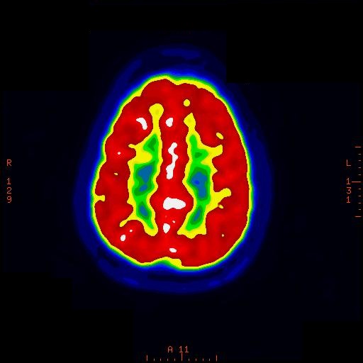
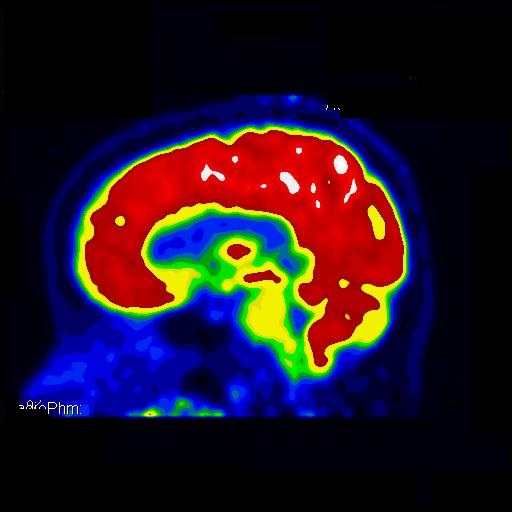
This is an example of a NORMAL PET CT scan. There is red coloring found along the surface of the brain indicating healthy blood flow to the underlying grey matter (neurons). The center of the brain has two large fluid filled chambers that contain cerebrospinal fluid (CSF). There is no blood flow to this region of the brain and therefore it appears blue in color. The underlying white matter (myelinated axons) which transport impulses from one neuron to another neuron appear green and yellow in color. They are green and yellow because they require very little blood flow to function.
PET CT
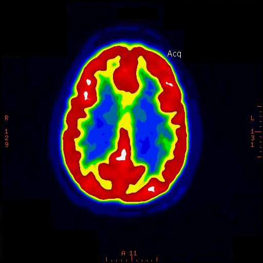

 RSS Feed
